Abstract
Introduction
This study evaluated and compared the shaping ability of the WaveOne Gold (Dentsply/Tulsa Dental Specialties, Tulsa, OK), TRUShape 3D Conforming File (Dentsply/Tulsa Dental Specialties), EdgeCoil (EdgeEndo, Albuquerque, NM), and XP-3D Shaper (Brasseler USA, Savannah, GA) endodontic file systems on oval-shaped canals using micro–computed tomographic (micro-CT) technology.
Methods
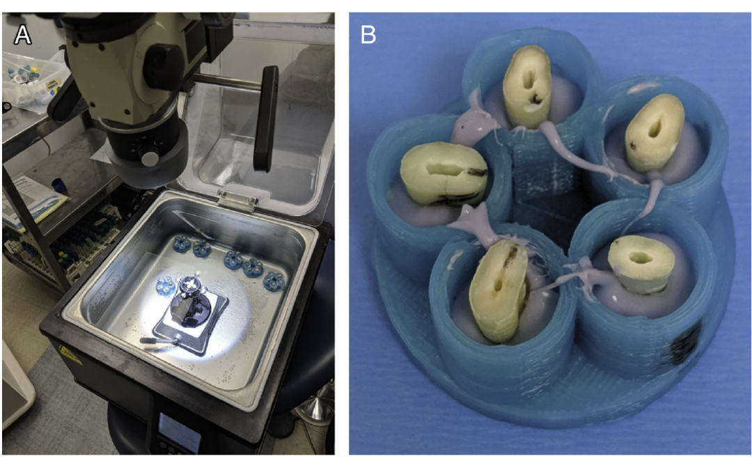
Thirty-two oval-shaped, single-canal extracted human teeth were decoronated 1 mm coronal to the cementoenamel junction and scanned via a micro-CT scanner (mCT100; Scanco Medical, Bassersdorf, Switzerland). Teeth were divided into 4 groups (n 5 8) and instrumented according to the manufacturer’s instructions. Coregistered images, before and after root canal preparation, were evaluated for morphometric measurements of the surface area, volume, structure model index (SMI), conicity, and percent of walls untouched using the manufacturer’s evaluation software (IPL Register, Scanco Medical). Data were statistically compared between groups using 1-way analysis of variance and within groups using a paired sample t test.
Results
Instrumentation with all file types increased the surface area, volume, and conicity between and within groups. There was no statistically significant difference between the groups for any of the rotary instruments used (P , .05).
Conclusions
Within the limitations of this study, it can be concluded that the WaveOne Gold, TRUShape, Edge Coil, and XP-3D Shaper file systems increase volume, surface area, and conicity of instrumented root canals. No file system was capable of contacting all of the surface area in any canal, but EdgeCoil attained the greatest increase in volume and surface area as well as the least amount of untreated voxels among all of the file systems tested.
Read Full Study Here: Micro–computed Tomographic Evaluation of the Shaping Ability of WaveOne Gold, TRUShape, EdgeCoil, and XP-3D Shaper Endodontic Files in Single, Oval-shaped Canals- An In Vitro Study